
Glaucoma Consults Round-up: When to treat narrow angles and too little too late.
/Wednesday October 14, 2009 and October 14, 2012
This is one of many articles that were corrupted in the migration from Squarespace 5 to 6 recently. I have manually re-entered the entire article as it still has some useful teaching points that were worth saving.
Case 1:
robschertzer #Glaucoma #Consult 58 yo WF told has angle closure needing iridotomy; narrow approach but wide open - no need for intervention.
Although not great evidence to support exactly when patients with narrow angles are at risk of having them close either acutely or gradually, patients are often referred to help determine whether they would benefit from a laser iridotomy to prevent angle closure.
The most obvious reasons to treat such patients is if they show signs or report symptoms of having had attacks of angle closure. Signs would include synechaie, areas of scarring between the peripheral iris and the trabecular meshwork drainage system. Symptoms would include episodes of headaches, especially if associated with eye pain or red eye. If either the signs or the symptoms are present, I tend to recommend treatment.
If there is no evidence of prior angle closure episodes, I tend to not recommend treatment, but often do recommend ongoing follow-up visits.
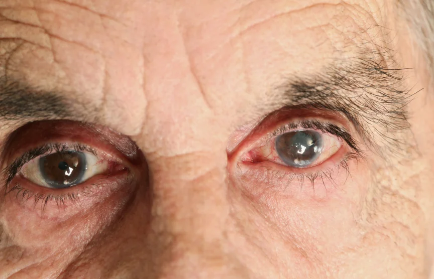
An important determining factor is the examination of the drainage channels using a mirrored (gonioscopy) lens. This overcomes total internal reflection of the patient's cornea to enable the image of those structures to be visualized, and not have their light rays bounce back into the eye because of the index of refraction of the cornea and its overlying tear film. This examination is best performed with an indentation type of gonioscopy lens so that the cornea can be gently indented during the examination in order to see if the angle position changes with gentle pressure. This process helps verify if there are any synechiae occluding the angle, suggesting prior episodes of angle closure.
Case 2:
robschertzer#Glaucoma #Consult 49 yo Asian M top half VF gone OU with IOP 25; longstanding COAG. Wish caught years earlier.14Oct2009 Case 2 HRT OU Report
14Oct2009 Case 2 HVF OS
14Oct2009 Case 2 HVF OD
This patient has been followed by the referring Optometrist for the past four years and is a high myope in the -9.00D range. There is a family history of cataracts and glaucoma in his mother. Visual acuity with correction is 6/7.5 OD and 6/12 OS. IOP readings are 25OU @1320hrs with pachymetry readings of 564 ums OD and 577 ums OS. His angles are wide open to the posterior Trabecular Meshwork. Both optic nerves show advanced glaucomatous damage along with peripapillary changes from myopia.
This is fairly tragic and one of the reasons we always hope for early detection and treatment of glaucoma. There is no way to really know when the glaucoma damage started, but it is likely to have been on-going for somewhere between 3-10 years. Perhaps the eye pressure was even higher at some point or fluctuates a great deal now as 25 alone would not result in rapid optic nerve damage. I base this educated guess on estimates that it takes up to 3 years from the onset of glaucoma until a defect can be confirmed on visual field testing and that it takes longer than that to document a change. This patient has advanced glaucoma damage and the best we can hope for now is to keep things from getting worse. Based on the Advanced Glaucoma Intervention Study, the goal of treatment would be to aim for an IOP reading always below 18 mmHg as that group in the study showed almost no change for the worse over 7 years of follow-up.
Case 3:
robschertzer #Glaucoma #Consult 53 yo WF hyperope, nasal step OS w/ increased cupping OS vs OD but still WNL. Follow for now.
This patient has been noted to have a repeatable Visual Field defect on Frequency Doubling Testing with IOP 22 OU and a +4.50 hyperope. Based on this single visit, it is difficult to confirm that the VF and disc appearance are actually related to glaucoma damage and may just be congenital. He warrants further follow-up to see if things change over time.
14Oct2009 Case 3 HRT OU Report
14Oct2009 Case 3 VF OS
14Oct2009 Case 3 VF OD
Case 4:
robschertzer #Glaucoma #Consult 83 yo Asian M from Taiwan; Tx'd x yrs Xalacom OD & Sancoba (B12) OS. Discs healthy, 1/3 angle recessed.
Little known 'fact' or perhaps fiction: Sancoba is a Japanese ophthalmic fomulation of Cyanocobalamin, Vit B12, that is used to help muscle function to prevent eyestrain. There is no such ocular formulation in North America and I would be interested in learning some science behind this use of Vitamin B12.
Case 5:
robschertzer#Glaucoma #Consult 44 yo Asian M, father/brother COAG, IOP 18 OU, increased 'cupping' OU (OD>OS) but normal VF. Follow for now.In addition to the family history of glaucoma, this patient was also noted by his referring Optometrist to have a disc haemmorhage in the right eye. In addition to those risk factors, this patient is also noted on examination to have pachymetry readings of 510ums OD and 511 OS. IOP readings were 18 OU at 1400hrs. Treat or not treat? What would you do with all these risk factors and normal Visual Fields?
Case 5 14Oct2009 HRT OS Stereometric
Case 5 14Oct2009 HRT OD Stereometric
Case 6:
robschertzer #Glaucoma #Consult 43 yo WM, uveitic glaucoma, debris in vitreous, optic nerve looks OK but IOP 30 on everything. Surgery?
This patient has chronic uveitis and has been followed out-of-town by an Ophthalmologist who does perform glaucoma surgery. Someone else had performed the cataract surgery on this patient in 1995 and this patient has been on topical steroids, drops and ointments since then. He first presented to the referring ophthalmologist with an IOP of 45 and is currently taking Combigan and Azopt for pressure control and Maxidex, PredForte and Nevanac for inflammation. Stopping the steroids altogether did not result in a lowering of his eye presssure so we cannot blame the pressure on his use of steroids, or at least the effect is no longer reversible. Perhaps out of desperation, Selective Laser Trabeculoplasty was even tried which did temporarily drop the pressure to the low 20's. I say out of desperation as we usually avoid laser interventions to the angle such as this in patients with inflammatory glaucomas as they can just increase inflammation without providing any pressure lowering.
Case 7:
robschertzer #Glaucoma #Consult 67 yo WM several abN FDT tests over the years but N on SAP; also has cataracts visually significant. Cat Sx booked.
Finished off the day with a patient with normal Standard Automated Perimetry despite prior abnormal Frequency Doubling Technology visual field tests. Patient was having trouble with normal reading activities with their current best spectacle correction. This patient is being booked for cataract surgery.